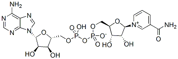
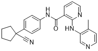
The metabotropic Coptisine-chloride glutamate receptor family, and the kainate/AMPA-type ionotropic glutamate receptor, are down-regulated in retinas from SIRT6 KO mice. Synaptic transmission between light-excited rod photoreceptors and downstream ON-bipolar neurons is indispensable for dim vision in the mammalian retina. Indeed, disruption of this process leads to congenital stationary night blindness in human patients. Notably, among the metabotropic receptors analyzed, Grm6 was found to be the most significantly down-regulated in KO retinas. Once released from the pre-synapsis, glutamate can bind to different glutamate receptors on the post-synapsis or can be removed from the synaptic cleft by high affinity glutamate transporters located on adjacent neurons and surrounding glial cells to prevent cell death. The major pathway of glutamate metabolism consists of glutamate uptake by glutamate transporters followed by enzymatic conversion of glutamate to non-toxic glutamine by glutamine synthetase. When a glutamate transporter is pharmacologically blocked, inner retinal neurons are exposed to a higher amount of endogenous glutamate, resulting in severe excitotoxic degeneration. These observations suggest that glutamate is neurotoxic when the uptake system is impaired rather than when the release is excessive. Alternatively, impaired expression of a post-synaptic receptor may contribute to the accumulation of the neurotransmitter in the inter-synaptic space. In this context, down-regulation of Grm6 could account for the increment of glutamate in the inter-synaptic space exerting a toxic effect that could explain the increase of TUNEL positive cellsfound in the inner nuclear layer of SIRT6 KO retinas. Since both bipolar and Mu��ller cells are involved in the generation of the b-wave, apoptotic cells in the inner nuclear layer would explain the alteration observed in the ERG. Overall, our studies demonstrated that SIRT6 deficiency causes major chromatin changes in the retina, which is accompanied by marked changes in expression of metabolic genes and metabotropic receptors, likely explaining the severe functional impairment observed in the SIRT6-KO retinas. Previous studies established sirtuins as critical Ascomycin modulators of metabolism, protecting against metabolic- and age-related diseases, such as diabetes, metabolic syndrome, cancer and neurodegenerative disorders. Our results  indicate that SIRT6 plays an important role in maintaining normal retinal function. Altered methylation patterns and histone modifications have been identified in different ocular diseases like diabetic retinopathy, glaucoma and age-related macular degeneration. However, a comprehensively characterized epigenomic signature for any ocular disease remains elusive. Several questions arise: is there any epigenetic mark that would predict the onset or the progression of an ocular disease? Do epigenetic factors regulate other cellular pathwaysthat may alter visual function? Although epigenetic therapies have proven to be effective in cancer applications, the benefits of these approaches have not yet been applied for human ophthalmic diseases. Thus far, there is no structural information available for any protein component of the MtrCDE tripartite complex system. However, it has been reported that individual protein components of this tripartite system are able to interact with each other, suggesting that the tripartite MtrCDE pump is assembled in the form of MtrD3-MtrC6-MtrE3.
Author: targets inhibitor
Given the importance of glucose availability for retinal function and the critical role of SIRT6
In the method developed here, proteins with different affinity of LZs localized to IBs were quantitatively analyzed in living cells using flow cytometry, while the E, K coil proteins in IB fractions was detected by electrophoretic methods after cell disruption in previous study. Therefore, the current method can be applied usefully for high throughput screening of PPI inhibitors, comparisons of interacting protein partners, and engineering binding affinities in bacterial cells. The mammalian retina is a highly metabolically active tissue and one of the most energy-consuming ones. It requires constant supply of blood glucose to sustain its function and its energy demand is normally met through the uptake of glucose and oxygen. Glucose movement across the blood�Cretinal barrier occurs mainly through the glucose transporter 1 and the need for glucose is evidenced by alterations in electroretinogramresponses and altered neurotransmitter release observed in hypoglycemic conditions. It has been shown that acute hypoglycemia decreases rod and cone vision, blurs central vision, and produces temporary central scotomas in humans. The consequence of sustained hypoglycemia on retinal function is less clear. Among many neuronal cellular events, action potential-mediated neuronal communication is believed to be a major process of energy consumption where energy cost comes mainly from postsynaptic receptor  activation. In the brain, most of the synaptic activity is mediated by glutamate, thus, the excitatory glutamatergic system represents the single largest energy user, consuming 50% of ATP in the brain. Photoreceptors convert light stimuli to electric impulses. Retinal ON bipolar cells receive direct glutamatergic input from photoreceptor cells. These cells exclusively express the class III G0-coupled type 6 metabotropic glutamate receptoras their primary postsynaptic glutamate receptor. Activation of mGluR6 initiates an intracellular signaling cascade ultimately leading to closure of cGMP gated cation channels and cell hyperpolarization. Thus, energy requirement and consumption in the retina changes greatly according to neuronal activity. Sirtuins are an evolutionarily conserved family of NAD + dependent deacylases that have been involved in many cellular responses to stress, including Evodiamine chromatin modifications, genomic stability, metabolism, inflammation, cellular senescence and organismal lifespan. In mammals, 7 sirtuin isoforms have been described that differ in their Kaempferide subcellular localization and substrates. It is currently accepted that sirtuins are crucial regulators of energy metabolism, likely through sensing changes in levels of intracellular NAD+. Among the members of this family of proteins, SIRT6 appears to have particular significance in regulating metabolism, DNA repair and lifespan. SIRT6 knockout mice appear normal at birth, but they rapidly develop a degenerative process that includes loss of subcutaneous fat, lymphopenia, osteopenia, and acute onset of hypoglycemia, leading to death in less than one month of age. Recently, Zhong et al. demonstrated that the lethal hypoglycemia exhibited by SIRT6 deficient mice is caused by an increased in glucose uptake in muscle and brown adipose tissue. At a molecular level, SIRT6 functions as a histone H3K9 and H3K56 deacetylase to control glucose homeostasis by inhibiting multiple glycolytic genes, including GLUT1, and by co-repressing Hif1a, a critical regulator of nutrient stress responses.
With a cellulose acetate dialyzer highlighting the potential deleterious effects associated with the dialysis treatment
We were unable to show a difference in serum T-BARswith short daily compared to conventional hemodialysis. Interestingly, phosphate has been shown to stimulate endothelial cell apoptosis which was characterized by increased oxidative stress. It is unclear if the effects of an increase in dialysis frequency and exposure to dialysis tubing and membranes is counterbalanced by the improvements in serum phosphate that have been reported with short daily dialysis. A generalized increase in inflammatory Epimedoside-A markers including IL-6 may occur via a number of mechanisms including volume overload and oxidative stress. We were unable to show a difference in IL-6 or albumin with short daily compared to conventional hemodialysis. In a prospective cohort study of 26 patients treated with short daily HD in-centre, Ayus et al reported significant improvements in hs-CRP values after 12 months. In a crosssectional study by Jefferies et al, lower CRP values were only seen for patients who were doing more frequent dialysis at home compared to those patients who were treated in-centre. Post dialysis sodium, ultrafiltration volumes and ultrafiltration rates were much lower for the patients being treated at home compared to the patients being treated in-centre suggesting that post-dialysis increases in serum sodium and/or interdialytic ECFV expansion may explain these differences. They were unable to demonstrate lower IL-6 levels between patients undergoing conventional and short daily dialysis in-centre who had similar post dialysis serum sodium levels and ultrafiltration rates. The impact of more frequent dialysis on markers of oxidative stress and inflammation requires further study. Our study has a number of limitations including the inability to blind the patients and investigators to the treatment group. We did not measure residual renal function and therefore could not assess the importance of this variable. However, the dialysis vintage of the patients suggests that many of them may have been anuric. Lastly, we did not measure ECFV at baseline and again at the end of the run-in phase; we are therefore unable to comment on the role changes in ECFV in the blood pressure improvement that was seen in the BP optimization phase. Strengths of our study over others that have been published to date include the run-in phase and use of standardized blood pressure algorithm. In summary after a 3-month run-in phase in which blood pressure was optimized by decreasing dialysate sodium, adjusting dry weight and anti-hypertensive medications, patients treated with short daily HD compared to conventional HD require fewer anti-hypertensive medications to achieve the same blood pressure. This effect on blood pressure control was not related to a reduction in ECFV, sympathetic nervous system activity, oxidative stress or inflammation. The mechanism by which short daily HD allows for decreased use of anti-hypertensive medication remains unclear but may be related to effects on sodium balance and changes in peripheral vascular resistance that require further study. It probably diverged from the primate orderabout 85 Procyanidin-B1 million years ago, which are now widely classified as a separate  taxonomic group of mammals. Consequently, tree shrew is a potentially useful animal model for some human diseases because of closer phylogenetic relationship with human.
In contrast to the general population urine volume are closely interrelated in severe
The association between urine osmolarity and the risk of initiating dialysis, a stronger marker for end stage renal disease, has not been previously investigated. Our data thus adds to the existing evidence. The data is in line with Torres et al. suggesting a faster renal function decline in patients with higher urine osmolarity. Our cohort appeared to have the highest urine osmolarity with a median of 510 mosm/L versus a mean of 3686159 mosm/L, and a mean of 270 to 334 mosm/L. It is therefore conceivable that lowering urine osmolarity below a specific threshold might be harmful to the kidney as well. A recent study addressing the association between sodium excretion, an important part of total urine osmolarity, and end-stage renal disease in patients with type 1 diabetes mellitus showed an inverse association with end stage renal disease. Furthermore, a U-shaped curve was described for sodium excretion and mortality, such that subjects with the highest and the lowest sodium excretion had the highest mortality, partly supporting the hypothesis of low urine osmolarity being associated with harmful effects. Interestingly, in the univariate versus the Gomisin-D multivariable analysis the strong effect of impaired renal function seems to obscure the positive correlation of osmolarity with the incidence of ESRD. Adjusting for creatinine clearance reveals this relationship. We conclude that among patients with equal renal function those with higher osmolarity values are more likely to progress to ESRD than those with lower osmolarity values in  our cohort. As described in the methods, Danshensu estimated urine osmolarity was calculated from urine sodium, potassium and urea concentrations in 24 hour urine samples. These are the dominant solutes in the urine. Other solutes represent less than 10% of total urinary solutes. Thus, estimated urine osmolarity is often used as an approximation of true osmolarity. Regarding the difference between osmolality and osmolarity, it is negligible in our study because the density of urine is very close to that of pure water in the range of values considered here. Other authors have used estimated urine osmolarity in several studies. Several authors have studied urine volume and GFR decline. Opposed to the prevailing view that water is beneficial in CKD, Hebert et al. reported higher GFR decline in higher urine volume quartiles. However, multivariable adjustment seemed to diminish the association. In support of Hebert et al., Wang et al. found a weak, but significant association between higher urine volume and GFR decline. A large study by Clark et al. showed a relationship between higher urine volume and lower rate of eGFR decline, which stayed significant after multivariable adjustment. Another large study reported worse kidney function in individuals with a lower self-reported fluid intake;however, neither urine volume nor urine osmolarity were evaluated. It has been suggested that lower mean baseline GFR in cohorts of Hebert et al. and Wang et al., which is associated with alterations in water metabolism, might explain these paradoxical findings. With falling GFR, the ability to concentrate urine to osmolalities greater than that of plasma is progressively lost.
Comparable urinary solute dilution varies considerably between individuals
Interestingly, median 24-hour urine osmolality is greater than that of plasma in humans, suggesting continuous antidiuretic action, which has been associated with renal function decline. Consequentially, Wang et al. recently devised a quantitative method to determine the amount of water needed on a case-by-case basis to achieve a mean urine osmolality Ginsenoside-Ro equivalent to that of plasma. Relationships between urine osmolarity and GFR decline have been described in two studies with contrasting results. We were interested in studying urine volume and urine osmolarity in terms of harder endpoints in chronic kidney disease. Thus we set out to study these variables in terms of risk of initiating dialysis, with death as a competing event. To describe intra- versus inter-individual variance of urine osmolarity, we conducted a variance component analysis including all run-in urine osmolarity values that were available for each patient using a mixed model with patients as levels of a random factor. The outcome variable was time to dialysis, with death as the competing event. Patients who were alive without dialysis at the time of their last visit were censored. Absolute event rates were computed as the number of events divided by the total follow-up time for all patients. Observations with missing values were not used in the calculated models. We described the distribution of time to dialysis using cumulative incidence functions, and compared groups using Gray��s test. Due to the established relationship between baseline creatinine clearance and risk of initiating dialysis/ESRD, and the known progressive loss in urine concentration ability with decreasing renal function, it seemed important to introduce creatinine clearance  as an Gentiopicrin adjustment factor in all further analyses. We fi ed two multivariate proportional sub-distribution hazards models for competing risk data according to Fine and Gray in order to assess the effect of urine osmolarity or volume on the risk for initiating dialysis. In these models, we considered osmolarity or urine volume and included those variables that either proved significant in a multivariate model or changed the log hazard ratio of osmolarity or urine volume by more than 15% when those variables were excluded from the analyses. We assumed that any variable not selected would have no relevant impact on our conclusions. All variables listed in Table 1 were considered as potential confounders. Results from multivariate competing risk regression were described by means of sub-distribution hazard ratios and 95% confidence intervals, and by computing and visualising estimated cumulative incidence curves at specific covariate values. As urine osmolarity and creatinine clearance were log-base-2 transformed, their SHR correspond to each doubling of these variables. We checked for significant pairwise interactions of variables and for time-dependent effects by including interactions with follow-up time. Non-linear effects were assessed by the method of fractional polynomials. For sensitivity analysis, we also estimated a cause-specific Cox regression model. The R-software, version 2.12, was used for statistical analysis. Other models were calculated for follow-up urine osmolarities and did stay significant after further adjustments.