Meta Description: Mechanistic analysis of a PEI-R848 nanovaccine platform revealing how TLR4 and NF-κB signaling, validated with AbMole inhibitors Resatorvid and PDTC, drives lymph node macrophage repolarization and amplifies antigen cross-presentation.
Target Keywords: nanovaccine, macrophage repolarization, TLR4, NF-κB, PEI, R848, lymph node, antigen cross-presentation, Resatorvid, PDTC, AbMole, innate immunity
Lymph nodes have always been viewed primarily through the lens of dendritic cell biology. Vaccine design overwhelmingly targets DC maturation, antigen cross-presentation, and subsequent T-cell priming, while macrophages residing in the same tissue are often treated as accessory cells at best, or as irrelevant bystanders at worst. A recent study in Acta Pharmaceutica Sinica B challenges this hierarchy by demonstrating that lymph node macrophages are not merely passive inhabitants but active gatekeepers whose phenotypic state dictates the amplitude of cellular immune responses. The work introduces a dual-adjuvant nanovaccine built from polyethyleneimine and resiquimod that simultaneously activates DCs and repolarizes macrophages, creating a coordinated innate immune environment that substantially amplifies antigen presentation.
The initial observation that prompted this approach came from comparing normal lymph nodes with tumor-draining lymph nodes. The latter displayed a striking accumulation of M2-like macrophages, reduced DC activation markers, and exhausted CD8+ T-cell populations. Depleting these macrophages with clodronate liposomes altered local immune dynamics, confirming that macrophage identity actively sculpts the lymph node microenvironment. This set up a clear premise: if macrophages in lymph nodes skew toward an immunosuppressive state, then any vaccine strategy ignoring them is operating with one hand tied behind its back.
The investigators turned to polyethyleneimine, a cationic polymer widely recognized for nucleic acid complexation but less appreciated for its innate immune activity. PEI activates TLR4, triggering downstream signaling through TRIF and MyD88 adapters. Resiquimod, a small-molecule TLR7/8 agonist, has established credentials in macrophage repolarization and DC activation. The critical question was whether these two agents would cooperate or merely additively stimulate. In bone marrow-derived DC cultures, PEI alone at low concentration failed to upregulate CD80 and CD86, yet when combined with R848, the proportion of activated DCs jumped from roughly 9% to 24%. At higher PEI concentrations, the combination produced a 3.1-fold increase over R848 alone and a 2.0-fold increase over PEI alone. This was not simple summation; it was genuine synergy.
The mechanistic basis for this synergy traced back to the TRAF3-TBK1-IRF3 axis. Both TLR4 and TLR7/8 signaling can feed into TRAF3, leading to TBK1 phosphorylation and subsequent IRF3/7 nuclear translocation to drive type I interferon production. Flow cytometry showed that p-TBK1 levels were markedly elevated in the combination group, and Western blotting confirmed robust IRF3 phosphorylation. Supernatants from combination-treated DCs induced substantially higher IFN-I reporter activity than either single agent, reaching approximately 5-fold above R848 alone and 7.6-fold above PEI alone. The implication is clear: concurrent engagement of TLR4 and TLR7/8 on DCs creates a feed-forward loop through shared adaptor proteins that amplifies innate immune activation beyond what either receptor achieves independently.
Where the study breaks genuinely new ground is in macrophage biology. BMDMs polarized to an M2 phenotype with IL-4 were exposed to PEI, R848, or both. The combination drove a pronounced shift toward M1-associated CD86 expression and elevated the M1/M2 ratio well beyond either monotherapy. More importantly, the combination triggered substantial secretion of IL-12 and IFN-β, cytokines that create a pro-inflammatory milieu capable of supporting downstream adaptive immunity. The magnitude was considerable: IL-12 in the combination group reached 2.1-fold higher than R848 alone and 2.9-fold higher than PEI alone.
To dissect the signaling architecture underlying this repolarization, the team employed two pharmacological inhibitors sourced from AbMole. Macrophages were pretreated with Resatorvid, a selective TLR4 inhibitor, at 100 nmol/L for one hour before PEI + R848 challenge. Resatorvid almost completely abolished the PEI + R848-induced increase in M1/M2 ratio, demonstrating that PEI-mediated TLR4 engagement is non-negotiable for phenotypic switching. Parallel experiments used PDTC, an NF-κB inhibitor, at 100 μmol/L with the same pretreatment protocol. PDTC significantly suppressed IL-12 secretion and blocked the repolarization response. Western blotting further showed that PEI + R848 strongly activated NF-κB, evidenced by increased p-NF-κB levels, and that this activation was sensitive to PDTC blockade. Collectively, the AbMole inhibitor studies established the TLR4/NF-κB axis as the dominant signaling conduit through which the dual-adjuvant system reprograms macrophage function.
This mechanistic clarity is worth emphasizing because it moves beyond phenomenology. Many adjuvant combinations produce impressive cytokine readouts without clear pathway attribution. Here, the use of Resatorvid and PDTC created a rigorous evidentiary chain: PEI engages TLR4, which signals through NF-κB, which drives both phenotypic conversion and IL-12 production. The inhibitors were not merely negative controls; they functioned as molecular scalpels that dissected the circuitry with precision.
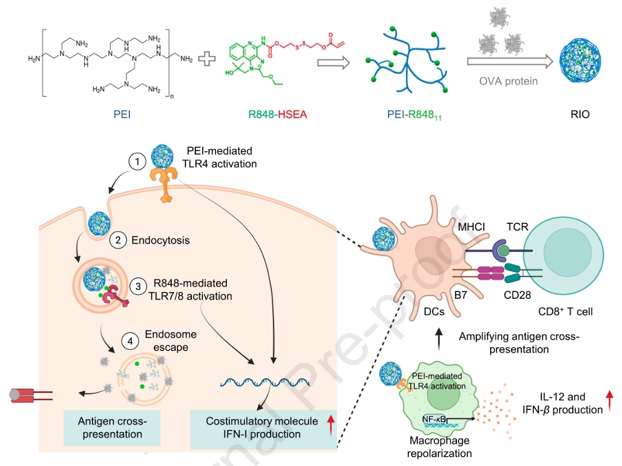
Building on these in vitro findings, the researchers constructed a nanovaccine by covalently conjugating R848 to PEI through a reducible disulfide linker, creating PEI-R848. The R848 prodrug was synthesized via reaction of triphosgene with HSEMA, followed by DMAP-catalyzed coupling to R848. Proton NMR confirmed successful conjugation, with characteristic peaks at 5.89–6.42 ppm verifying the acrylate incorporation. Two grafting densities were prepared—11 and 6 R848 molecules per PEI chain—with the higher density variant proving superior for antigen delivery.
The hydrophobic R848 moieties transformed PEI from a simple cationic polymer into an amphiphilic construct capable of self-assembling with ovalbumin into discrete nanoparticles. Agarose gel electrophoresis showed that PEI-R848 completely retarded OVA migration at 2:1, 1:1, and 1:2 mass ratios, whereas unmodified PEI left substantial free antigen. Encapsulation efficiency exceeded 90% across all ratios. Dynamic light scattering and transmission electron microscopy revealed uniform spherical particles of approximately 221 nm at the 2:1 ratio, with zeta potentials around +32.5 mV. Stability in serum-containing medium was best at the 2:1 ratio, where particles remained monodisperse over seven days; higher antigen loads produced aggregates. The disulfide linker proved functionally relevant—R848 release reached 85% at 24 hours in 10 mmol/L glutathione but remained below 20% in its absence, ensuring that adjuvant activation occurs primarily intracellularly.
Intracellular trafficking studies in DCs showed robust cytoplasmic FITC-OVA delivery with PEI-R848, with confocal microscopy revealing substantial antigen escape from lysosomal compartments. This endosomal escape is mechanistically important because cytosolic antigen access is required for MHC-I cross-presentation. Indeed, surface SIINFEKL-H-2Kb complexes on DCs reached nearly 7% with RIO at the 2:1 ratio, a 6-fold improvement over free antigen. Co-culture with OT-I CD8+ T cells confirmed that this enhanced cross-presentation translated into vigorous T-cell proliferation.
In vivo lymph node targeting experiments showed that RIO nanoparticles accumulated substantially better than free antigen after subcutaneous administration. Flow cytometry of inguinal lymph nodes revealed significantly higher OVA uptake by DCs in the RIO group. Critically, when macrophages were depleted with clodronate liposomes prior to vaccination, both DC activation and antigen presentation dropped markedly. This macrophage dependency is the central conceptual advance: the nanovaccine does not simply bypass macrophages to reach DCs; it actively converts macrophages into collaborators that secrete IL-12 and IFN-β, which in turn amplify DC function and T-cell priming.
The phenotypic shift in lymph node macrophages was confirmed directly—RIO treatment increased CD80+ M1-like macrophages and reduced CD206+ M2-like populations compared to physical mixtures of the components. By Day 7 post-immunization, splenic tetramer analysis showed 2.3-fold more antigen-specific CD8+ T cells in RIO-immunized animals than in free antigen recipients, and IFN-γ ELISpot counts were correspondingly elevated.
What makes this platform architecturally elegant is the integration of structural and immunological functions into a single molecular design. PEI provides cationic charge for antigen complexation and TLR4 activation; R848 contributes hydrophobicity for self-assembly and TLR7/8 agonism for endosomal innate immune sensing; the disulfide linker ensures redox-triggered adjuvant release. No single component is expendable, and the synergy between TLR4 and TLR7/8 signaling—validated through the AbMole inhibitor studies—provides a rational basis for why the integrated nanovaccine outperforms simple physical mixtures.
For immunologists and vaccine engineers, this work carries several actionable insights. First, it elevates lymph node macrophages from background noise to legitimate targets for vaccine adjuvantation. Second, it demonstrates that TLR pathway combinations can be rationally designed rather than empirically screened, provided the signaling architecture is mapped with appropriate pharmacological tools. Third, it highlights the value of reducible linkers in controlling adjuvant bioavailability, ensuring that innate immune activation is concentrated where antigen processing occurs.
In sum, the study constructs a coherent mechanistic narrative in which a PEI-R848 nanoplatform engages both DCs and macrophages in lymph nodes, with the latter requiring TLR4/NF-κB signaling to switch from immunosuppressive to immunostimulatory phenotypes. The rigorous pathway validation, enabled by the TLR4 inhibitor Resatorvid and the NF-κB inhibitor PDTC from AbMole, transforms what could have been a purely descriptive adjuvant study into a precisely mapped immunological circuit. Future iterations of this platform will likely explore alternative antigen cargoes and further dissect the kinetics of macrophage-DC crosstalk, but the foundational principle is now established: effective cellular immunity demands coordinated reprogramming of both professional antigen-presenting cells and the macrophage networks that regulate their microenvironment.
AbMole Product Integration in This Study
Product 1: Resatorvid (AbMole Biotechnology, Houston, TX, USA)
Application: Selective TLR4 inhibitor for mechanistic validation of PEI-mediated macrophage repolarization.
Experimental Details:
-
Concentration: 100 nmol/L
-
Protocol: Bone marrow-derived macrophages (BMDMs) were polarized to M2 phenotype with IL-4 for 24 h, then pretreated with Resatorvid in serum-free medium for 1 h prior to PEI + R848 stimulation.
-
Context: Used alongside PEI (5 μg/mL) and R848 (1 μg/mL) co-incubation for an additional 24 h; each condition performed in triplicate.
Key Findings Enabled by Resatorvid:
-
Resatorvid pretreatment markedly suppressed PEI + R848-induced macrophage repolarization toward pro-inflammatory phenotype.
-
The M1/M2 ratio increase driven by PEI + R848 was significantly attenuated, confirming that PEI-mediated TLR4 activation is obligatory for phenotypic switching.
-
IL-12 secretion induced by the dual-adjuvant combination was also dampened, positioning TLR4 signaling upstream of cytokine production.
Product 2: PDTC (AbMole Biotechnology, Houston, TX, USA)
Application: NF-κB pathway inhibitor for dissecting downstream signaling in macrophage functional reprogramming.
Experimental Details:
-
Concentration: 100 μmol/L
-
Protocol: Identical pretreatment workflow as Resatorvid—1 h pre-incubation in serum-free medium before PEI + R848 challenge.
-
Readouts: Flow cytometric analysis of macrophage phenotypes and ELISA quantification of IL-12 in culture supernatants.
Key Findings Enabled by PDTC:
-
PDTC pretreatment significantly blunted IL-12 secretion triggered by PEI + R848.
-
Western blot analysis confirmed that PEI + R848 robustly activated NF-κB signaling (elevated p-NF-κB), and PDTC blockade suppressed this response.
-
Together with Resatorvid data, the AbMole inhibitors established the TLR4/NF-κB axis as the critical signaling route through which the dual-adjuvant system rewires macrophage function.