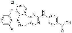
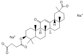
In addition to its well-known fibrinolytic properties, this protease has also been reported to play a critical role in various other physiological and pathophysiological processes including angiogenesis, wound healing, and inflammation. In this context, plasmin is suggested to initiate intracellular signaling pathways as well as to activate extracellular matrix degrading enzymes ultimately facilitating cell adhesion and migration. Despite recent concerns about the safety of the broad-spectrum serine protease inhibitor aprotinin, clinical trials revealed beneficial effects of this naturally occurring substance for the prevention of postischemic organ dysfunction. Here, aprotinin has been suggested to suppress the transcription of genes which have been implicated in the evolution of the postischemic inflammatory response. The consequences for each single step of the leukocyte recruitment process during I/R, however, have not yet been studied. Previous studies have implicated the serine protease plasmin as well as plasminogen activators in the regulation of leukocyte migration to the site of inflammation. Interestingly, lysine analogues such as tranexamic acid or e-aminocaproic acid have recently been reported to effectively and safely inhibit plasmin activity. The effect of these synthetic plasmin inhibitors on postischemic leukocyte responses has not yet been evaluated. In the early reperfusion phase, remodeling processes within the perivenular basement membrane have been described which are thought to compromise microvascular integrity and to pave the way for the excessive leukocyte infiltration of reperfused tissue. Due to its capability to disintegrate components of the venular basement membrane as well as to activate other ECMdegrading proteases, plasmin has been implicated in these events. The effect of plasmin inhibitors and aprotinin on remodeling processes within the postischemic vessel wall has not yet been investigated. Restoration of blood flow is the overall goal for successful organ transplantation as well as for the treatment of myocardial infarction, hemorrhagic shock, and stroke. As a consequence of this inevitable approach, however, neutrophils accumulate within the postischemic microvasculature and compromise reperfusion of the affected organ. Subsequently, transmigrating neutrophils release reactive oxygen species, cytokines, and proteases, impairing microvascular integrity and promoting postischemic tissue injury. Notably, PF-04217903 extravasated neutrophils also contribute to tissue healing and regeneration collectively emphasizing neutrophil recruitment as a key event in the pathogenesis of I/R injury. Using different animal models, the serine protease plasmin as well as plasmin activators have been implicated particularly in the migration of monocytes, but also in the recruitment of neutrophils. Moreover, clinical trials revealed beneficial effects of the broad-spectrum serine protease inhibitor aprotinin for the prevention of postischemic organ dysfunction after coronary revascularization. In this context, aprotinin has been  reported to suppress the transcription of genes which are supposed to play a major role in the postischemic inflammatory response. The resulting consequences for each single step of the leukocyte recruitment process, however, remained LDN-193189 ALK inhibitor unclear. Using near-infrared RLOT in vivo microscopy on the mouse cremaster muscle, we systematically analyzed the effects on postischemic rolling, firm adherence, and transmigration of leukocytes of the broad-spectrum serine protease inhibitor aprotinin, a naturally occurring bovine protein.
Month: August 2019
Due to an enhancement of COX-2 protein the form of chemical descriptors computable numerical attributes in vector form
Numerous molecular 3D-descriptors and alignment methods have been proposed. Examples include CoMFA, Randic molecular profiles, 3DMoRSE code, invariant MK-0683 moments and radial scanning and integration, radial distribution function descriptors, WHIM, length-to-breadth ratios, USR, ROCS, VolSurf, GETAWAY, and shrinkwrap surfaces, to name just a few prominent representatives. In computer MK-4827 graphics, several methods exist for the more general problem of comparing arbitrary 3D objects, including distribution-based shape histograms, the D2 shape descriptor, and, the scaling index method; the viewbased methods of extended Gaussian images, and the light field descriptor; the surface decomposition-based methods -Licarin-B-chemical-structure.gif) of Zernike moments, REXT, and spherical harmonics descriptors. Spherical harmonics have been used in cheminformatics as a global feature-based parametrization method of molecular shape. Their attractive properties with regard to rotations make them an intuitive and convenient choice as basis functions when searching in a rotational space. A review article by Venkatraman et al. highlights applications of spherical harmonics to protein structure comparison, ligand binding site similarity, protein-protein docking, and virtual screening. Jakobi et al. use spherical harmonics in their ParaFrag approach to derive 3D pharmacophores of molecular fragments. Recently, Ritchie and co-workers have applied the ParaSurf and ParaFit methodologies in a virtual screening study on the directory of useful decoys data set, which motivates 3D shape-property combinations specifically for flexible ligands. The DUD data set was also used in a comparative analysis of the performance of various shape descriptors alone and in combination with property and pharmacophore features. See the section on related methods for further discussion of spherical harmonics approaches. In this work, we introduce a partially rotation-invariant descriptor of molecular shape based on spherical harmonics decomposition coefficients. The idea is to decompose the molecular surface using spherical harmonics and to use the norm of the decomposition coefficients as a description of molecular shape. In this, we take advantage of the fact that the norm of the coefficients does not change under rotation around the z-axis, which we align to the primary axis of the molecule. We retrospectively evaluate our descriptor, and prospectively apply it to screen for novel inhibitors of the enzymes cyclooxygenase-1 and cyclooxygenase-2. Particular focus is on the practical application of the virtual screening technique as an evaluation of its actual suitability for early-phase drug discovery. The inhibitory data obtained from the whole blood assay might be meaningful for further hit optimization. Compounds that are active in this assay are not snatched away by binding to serum albumin, but cross the cell membrane and overcome possible interactions with cellular substances or enzymes. This could explain why compounds 5 and 9 are active in the enzyme assay, but inactive in the whole blood assay. In contrast, compounds 6,10,2and 8, which were moreactive in the whole blood assay, possibly interact with the arachidonic acid pathway in other ways than direct inhibition of COX-1 or COX-2. Also, these compounds might be metabolized by cellular enzymes to more active derivatives, but this hypothesis needs to be tested by further experiments. Compound 8 is of special interest, as it induces PGE2 production up to 322%. This increase could be due to an activation of enzyme activity, possibly by binding to the “inactive” monomer of the COX-homodimer complex.
of Zernike moments, REXT, and spherical harmonics descriptors. Spherical harmonics have been used in cheminformatics as a global feature-based parametrization method of molecular shape. Their attractive properties with regard to rotations make them an intuitive and convenient choice as basis functions when searching in a rotational space. A review article by Venkatraman et al. highlights applications of spherical harmonics to protein structure comparison, ligand binding site similarity, protein-protein docking, and virtual screening. Jakobi et al. use spherical harmonics in their ParaFrag approach to derive 3D pharmacophores of molecular fragments. Recently, Ritchie and co-workers have applied the ParaSurf and ParaFit methodologies in a virtual screening study on the directory of useful decoys data set, which motivates 3D shape-property combinations specifically for flexible ligands. The DUD data set was also used in a comparative analysis of the performance of various shape descriptors alone and in combination with property and pharmacophore features. See the section on related methods for further discussion of spherical harmonics approaches. In this work, we introduce a partially rotation-invariant descriptor of molecular shape based on spherical harmonics decomposition coefficients. The idea is to decompose the molecular surface using spherical harmonics and to use the norm of the decomposition coefficients as a description of molecular shape. In this, we take advantage of the fact that the norm of the coefficients does not change under rotation around the z-axis, which we align to the primary axis of the molecule. We retrospectively evaluate our descriptor, and prospectively apply it to screen for novel inhibitors of the enzymes cyclooxygenase-1 and cyclooxygenase-2. Particular focus is on the practical application of the virtual screening technique as an evaluation of its actual suitability for early-phase drug discovery. The inhibitory data obtained from the whole blood assay might be meaningful for further hit optimization. Compounds that are active in this assay are not snatched away by binding to serum albumin, but cross the cell membrane and overcome possible interactions with cellular substances or enzymes. This could explain why compounds 5 and 9 are active in the enzyme assay, but inactive in the whole blood assay. In contrast, compounds 6,10,2and 8, which were moreactive in the whole blood assay, possibly interact with the arachidonic acid pathway in other ways than direct inhibition of COX-1 or COX-2. Also, these compounds might be metabolized by cellular enzymes to more active derivatives, but this hypothesis needs to be tested by further experiments. Compound 8 is of special interest, as it induces PGE2 production up to 322%. This increase could be due to an activation of enzyme activity, possibly by binding to the “inactive” monomer of the COX-homodimer complex.
BH3-only proteins antagonize the pro-survival function of Bcl-2 proteins or may activate
The concept of developing target-specific drugs for treatment of cancer has not been as successful as initially envisioned. The success rate of oncology drugs from first-in-man to registration during 1991�C2000 was only around 5% for 10 major high content screening pharma companies. A major causes of attrition in the clinic is lack of drug efficacy. This realization has lead to a renewed interest in the use of bioassays for drug development in the field of oncology. One attractive screening endpoint is apoptosis since this form of cell death is induced by many clinically used anticancer agents. Natural products have been used as source of novel therapeutics for many years. Natural products have been selected during evolution to interact with biological targets and their high degree of chemical diversity make them attractive as lead structures for discovery of new drugs. A number of plant-derived anticancer drugs have received FDA approval for marketing: taxol, vinblastine, vincristine, topotecan, irinotecan, etoposide and teniposide. Antibiotics from Streptomyces species, including  bleomycins, dactinomycin, mitomycin, and the anthracyclines daunomycin and doxorubicin are important anticancer agents. More recently developed anticancer agents such as the Hsp90 inhibitor geldanamycin was also isolated from Streptomyces. Marine organisms have also been used as source for the search of anticancer agents. Interesting compounds, including bryostatin, Afatinib ecteinascidin and dolastatin, have been identified. Although being the source of lead compounds for the majority of anticancer drugs approved by the Food and Drug Administration, natural products have largely been excluded from modern screening programs. We here used a high-throughput method for apoptosis detection to screen a library of natural compounds using a human colon carcinoma cell line as screening target. One of the most interesting hits in this screen was thaspine, an alkaloid from the cortex of the South American tree Croton lechleri. We show that thaspine is a topoisomerase inhibitor which is active on cells overexpressing drug efflux transporters. Thaspine has previously been described to have anti-tumor activity in the mouse S180 sarcoma model. To examine whether in vivo anti-tumor activity is associated with induction of apoptosis, SCID mice carrying HCT116 xenografts were treated with thaspine and tumor sections were stained with an antibody to active caspase-3. Positivity was observed in tumor tissue at 48 hours after treatment with 10 mg/kg thaspine. We also utilized caspase-cleaved CK18 as a plasma biomarker for tumor apoptosis. When applied to human xenografts transplanted to mice, this method allows determination of tumor apoptosis independently of host toxicity. Most forms of cancer therapeutics induce the mitochondrial pathway of apoptosis. This pathway is associated with opening of the mitochondrial permeability transition pore. We examined whether thaspine induced a decrease in HCT116 mitochondrial membrane potential using the fluorescent probe tetramethyl-rhodamine ethyl ester. Mitochondria in thaspine-treated cells underwent a shift to lower DyM values. A hallmark of the mitochondrial apoptosis pathway is release of cytochrome c from mitochondria to the cytosol. Thaspine was found to induce a decrease in the levels of mitochondrial cytochrome c and an increase of the levels in the cytosol. The Bcl-2 family proteins Bak and Bax are key regulators of the mitochondrial apoptosis pathway. During apoptosis, the conformation of these proteins is altered. Experiments using conformation-specific antibodies showed that thaspine induce conformational activation of both Bak and Bax.
The emergence of resistance and residual disease can eventually lead to progression of CML despite treatments
Imatinib, dasatinib or nilotinib resistance may emerge through point mutations in Bcr-Abl, Bcr-Abl gene amplification and/or an increase in Bcr-Abl protein levels. To investigate alternative treatments for these particular cases, we have indeed developed two different cell lines derived from K562 and LAMA84 cell lines, which are completely resistant to 1��M imatinib. While the levels of Bcr-Abl and P-Bcr-Abl in LAMA84-R are much AB1010 higher than in LAMA84-S cells, the levels of Bcr-Abl and P-Bcr-Abl in K562-R compared with K562-S are much closer to each other. Thus, the increased expression of Bcr-Abl is probably at least in part responsible for the LAMA-R resistance to imatinib, dasatinib and nilotinib, while possible mutations may be responsible for the K562-R resistance. Additionally, we have used the Baf3 Bcr-Abl T315I cell line, derived from Baf3, which is also resistant to 1��M imatinib and at least partially resistant to dasatinib and nilotinib treatments. In addition to its effect on imatinib-sensitive cell lines, the bortezomib/paclitaxel regimen was able to induce caspase cleavage, a measure of caspase activation, in K562-R cells and significant downregulation of the total levels and phosphorylation of Bcr-Abl in all tested TKIs-resistant cell lines. Thus, such combination may be a good strategy to treat resistant cases due to either an increase in Bcr-Abl expression or Bcr-Abl mutations that abrogate imatinib, dasatinib or nilotinib inhibitory effects. Notably, in addition to the bortezomib/paclitaxel regimen, our results demonstrate that bortezomib, in combination with other mitotic inhibitors that act by inducing mitotic arrest through various mechanisms, inhibits Bcr-Abl and results in caspase 3 activation. It has previously been established that inhibition of Bcr-Abl or knock-down of Bcr-Abl induces caspase activation and apoptosis. Thus, our results indicate that Bcr-Abl down-modulation contributes, at least in part, to caspase activation and induction of cell death. Both docetaxel and vincristine are FDA-approved for the treatment of several malignancies, alone or in combination. Interestingly, a recent study concluded that BI 2536 has growth inhibitory effects on Bcr-Abl-positive cells that are not amplified by bortezomib after 16h of co-treatment. In contrast, we are showing here that the combined treatment of bortezomib 9nM with BI 2536 8nM for 60h is significantly more effective in inducing caspase activation, PARP cleavage and cell death compared with single treatments, in both K562 and K562-R cells. The longer time needed for bortezomib to amplify the effects of BI 2536 might be explained by the involvement of transcriptional mechanisms in bortezomib/BI 2536-induced cell death, although further experiments are needed to clarify this aspect. Recently, two other drugs were approved by FDA for the treatment of patients with CML whose tumors are resistant to or who cannot tolerate Imatinib, Dasatinib or Nilotinib therapies: bosulif and synribo. Bosutinib, approved on September 4, 2012, is a TKI inhibitor efficient against many Bcr-Abl mutations, except T315I. Omacetaxine mepesuccinate, approved on October 26, 2012, is a non-TKI drug intended to be used when leukemia progresses after therapy with at least two TKIs. While the drug can be used for the treatment of CML patients  with T315I mutation, it shows significant hematologic toxicity in clinical trials: thrombocytopenia, neutropenia, and anemia. While these two new approved drugs offer an option for many patients with imatinib, dasatinib and nilotinibresistant CML, novel better strategies have to be developed. In contrast with bosutinib, our combined treatment with bortezomib and mitotic inhibitors is able to target Bcr-Abl with T315I mutation. Moreover, lower concentrations of each drug can be used in synergistic combinations, which may reduce toxicity. However, the toxicity of our MLN4924 regimens remains to be established.
The combined treatment was highly effective in decreasing the total levels and phosphorylation of Bcr-Abl T315I
Our results show that bortezomib and paclitaxel combined treatment is able to target the TKIsresistant cell lines with the T315I mutation in Bcr-Abl. Collectively, our findings indicate that the bortezomib in combination with four different mitotic inhibitors, that repress mitosis by different mechanisms are able to shut down Bcr-Abl activity and result in caspase-dependent cell death in TKIs-resistant and -sensitive Bcr-Abl-positive cell lines. A schematic representation of these findings is presented in Figure 7. Our results Screening Libraries demonstrate that regimens of bortezomib combined with mitotic inhibitors are associated with Bcr-Abl and/or P-BcrAbl downregulation. Few other agents have been shown to induce a significant Bcr-Abl downregulation when used in combination with imatinib. Moreover, the pan-CDK PCI-32765 inhibitor flavopiridol, the heat shock protein 90 antagonist 17-AAG and the histone deacetylase inhibitor SAHA were previously revealed to induce apoptosis in combination with bortezomib, an effect associated with Bcr-Abl downregulation. Although the exact mechanism of Bcr-Abl downregulation is still unclear, it seems plausible that the decrease of Bcr-Abl levels and its inactivation contribute, at least in part, to the caspase-mediated cell death induced by these combinations, including the bortezomib/mitotic inhibitors regimens. Our results point out that a bortezomib/paclitaxel combination inhibits STAT3 and STAT5 activation. Bortezomib/BI 2536 combination similarly results in a decrease in P-STAT5 levels in K562 cells. As previously shown, Bcr-Abl phosphorylates and activates STAT3 and STAT5 transcription factors resulting in cellular survival and proliferation. Constitutive activation of STAT5 is known to be critical for the maintenance of chronic myeloid leukemia and STAT3 is also constitutively active in Bcr-Abl-positive embryonic stem cells. Thus, cell death induced by inhibition of Bcr-Abl with imatinib in Bcr-Abl-positive cells  is at least in part related to the inhibition of STAT signaling. Additionally, it is known that JAKSTAT pathway activation contributes to imatinib and nilotinib resistance in Bcr-Abl-positive progenitors. All these findings suggest that STAT3/STAT5 signaling inhibition plays an important role in bortezomib/paclitaxel- or bortezomib/BI 2536-induced cell death, in Bcr-Abl-positive cells. Several pathways are known to be critical downstream mediators of the Bcr-Abl pro-survival and pro-leukemogenic effects. Bcr-Abl is phosphorylated at multiple phosphorylation sites, resulting in binding/phosphorylation of downstream BcrAbl mediators. Phosphorylation of Tyrosine 177 induces the formation of a Lyn – Gab2 – Bcr-Abl complex, important in BcrAbl-induced tumorigenesis. Lyn tyrosine kinase binding to phosphorylated and active Bcr-Abl leads to Lyn activation by phosphorylation. Lyn further regulates survival and responsiveness of CML cells to inhibition of Bcr-Abl kinase. Interestingly, Lyn kinase can also phosphorylate Bcr-Abl, resulting in a potential feedback mechanism. Additionally, Bcr-Abl phosphorylates CrkL adaptor protein, an event needed for Bcr-Abl-induced leukemia. CrkL can enhance cell migration and Bcr-Abl-mediated leukemogenesis. Thus, Lyn and CrkL are key regulators and downstream mediators of BcrAbl-induced survival and leukemogenesis that can be inhibited by downregulation or inhibition of Bcr-Abl. Our results demonstrate that the combined treatment with bortezomib and paclitaxel is able to inhibit the activity of these important BcrAbl downstream mediators. JNK activation was previously associated with apoptosis induced by bortezomib in Bcr-Abl-positive cells and by bortezomib in combination with the pan-CDK inhibitor Flavopiridol in both Bcr-Abl-positive and negative leukemic cells. In addition, several other studies pointed out the role of JNK activation in cell death of Bcr-Abl-positive or -negative cells. Thus, the activation of JNK seen in our results following bortezomib/paclitaxel treatment in Bcr-Abl-positive cells may contribute to cell death.