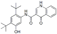
This process leads to the activation of each receptors’ tyrosine kinase and the subsequent phosphorylation of tyrosine residues located on their C-terminal tails. Phosphorylated tyrosine residues recruit various intracellular adaptor and effector molecules that result in the propagation of growth promoting signal transduction cascades. Membrane-bound HER receptors WY 14643 activate numerous tumor promoting signaling cascades via this mechanism, including the PI3K/AKT, Ras/Raf/Mek/Erk, PLCc/PKC, and signal transducer and activator of transcription pathways. While the classical membrane-bound functions of HER family RTKs have been extensively studied, accumulating data suggest that these receptors can be found in the cell��s nucleus where they can function as co-transcriptional activators. To date, EGFR, HER2, and a nuclear variant of HER3 have been shown to function as co-transcriptional activators for cyclin D1. Clinically, nuclear EGFR has been correlated with poor overall survival in breast, ovarian, oropharyngeal, and gallbladder cancers. Nuclear EGFR has also been shown to play a role in resistance to numerous cancer therapies, including radiation, cisplatin, and the anti-EGFR therapies cetuximab and gefitinib. Collectively, these pivotal studies suggest that nuclear HER family receptors may enhance the tumorigenic phenotype of cancer cells, and therefore their nuclear roles must be further elucidated. The HER3 receptor has recently come to the forefront as playing a key role in HER family driven cancers. HER3 is a unique HER family member in that it has diminished tyrosine kinase activity due to the lack of specific amino acids within its kinase domain. However, HER3 plays a crucial role as an allosteric activator of other HER family members in addition to functioning as a signaling substrate through the direct recruitment and activation of PI3K. With the discovery of the various functions of nuclear EGFR and HER2, recent interest has prompted the investigation of nuclear HER3. HER3 was first identified to be nuclear localized in normal and malignant MG132 Proteasome inhibitor mammary epithelial cells in 2002. HER3 was also shown to be prominently nuclear localized in malignant prostate cancer tissues, where it was correlated with risk of disease progression. Most recently, two nuclear C-terminal splice variants of HER3 were identified and shown to function as co-transcriptional activators. Thus, the functions of nuclear HER3 are just beginning to unfold. In the current study we focused on identifying the amino acid regions on the C-terminal tail of HER3 that function as transactivation domains. First, numerous cancer cell lines were characterized for the expression of HER3, which was prominently nuclear localized in its full-length form. Next, various HER3 intracellular cytoplasmic domain regions fused to  the Gal4 DNA binding domain were analyzed, and the Cterminal domain of HER3 was shown to activate the Gal4 upstream activation sequence fused to luciferase. To identify specific C-terminal TADs, we created nine HER3-CTD truncation mutants and identified a bipartite region of 34 and 27 amino acids in length that contained the majority of HER3��s transactivation potential. To determine if the identified B1 and B2 TADs could augment nuclear HER3’s transcriptional function in cells, we first identified that full-length nuclear HER3 could associate and activate a 122 bp region of the cyclin D1 promoter when overexpressed. Importantly, the overexpression of HER3 lacking the B1 and B2 regions was severely hindered both in activating the 122 bp region of the cyclin D1 promoter and in enhancing transcription from the endogenous cyclin D1 gene. Collectively, these data suggest that this bipartite region of HER3 functions as a prominent TAD to mediate HER3’s nuclear functions.