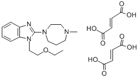
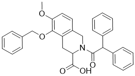
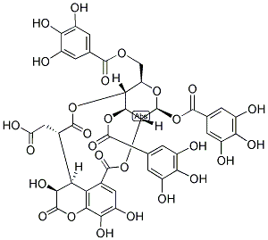
One study did examine nasal AEC cultured from 15 infants. However, the youngest infant was a month old, they were ‘held tightly by a nurse’ and because they were sedated with paracetamol and rectal midazolam they had to be monitored for 8 hours. This study also reported a single episode of epistaxis in their 15 subjects. In contrast, we observed no epistaxis in 155 neonates, this being most AbMole Aristolochic-acid-A likely due to the smaller diameter of our sampling brush, compared with the 5.5 mm brush used in the previous study. The paucity of erythrocytes identified in the cytospin preparations would suggest that the sampling procedure is relatively atraumatic in our hands. Our AbMole Niflumic acid methodology for sampling nasal AEC by nasal brushing in non-sedated neonates could prove a useful research tool and is especially applicable to large longitudinal studies. In addition to investigating the effect of in-utero influences on AEC phenotype, procuring neonatal airway epithelial tissue for culture has a variety of potential applications. As well as functional studies in submerged monolayers, it is likely that that our method could be adapted to grow neonatal nasal AEC in air-liquid interface cultures, which are thought to be more representative of conditions in vivo, this work is underway. Furthermore, obtaining nasal airway epithelial cells shortly after birth represents a privileged source of “na? ��ve” tissue not yet exposed to the modifying effects of post natal environmental pollutants and pathogens. As such, the tissue specific epigenetic effects of such exposures throughout life on the expression of  genes involved in inflammatory mediator release could be explored. Based on our previous success culturing nasal airway epithelial cells from both adults and children, a study investigating repeated isolation of nasal cells from the same individual over time would be feasible. If neonatal nasal AEC are shown to be associated with subsequent asthma/allergic rhinitis and recognized/putative risk factors for these conditions, it may be possible to use neonatal nasal AEC as a neonatal biomarker in antenatal intervention studies. In the clinical setting it may be possible to use neonatal nasal AEC as a predictive biomarker to quantify subsequent risk of asthma. Of particular importance will be to confirm the hypothesis that the AEC of neonates who subsequently develop childhood asthma differ from AEC of children who do not develop asthma. In addition it will be important to ascertain whether neonatal AEC function is associated with established risk factors for childhood asthma, e.g. parental asthma, maternal smoking and maternal diet. Indeed we are currently using the methodology developed and reported here in an ongoing birth cohort study that is aimed at addressing some of these issues.
Month: April 2019
RAGE has been linked to several chronic diseases the progression of diabetic vascular complications
In hyperglycemic conditions, levels of precursors of triose phosphate, such as glucose or fructose, are increased. After nonenzymatic fragmentation, high serum levels of MGO were observed in patients with either type 1 or type 2 diabetes. MGO as one of the most reactive dicarbonyls, is considered to be an important glycating agent  to consider for glycation damage to the mitochondrial proteome. Moreover, the cytotoxicity of MGO is mediated by the modification of deoxyribonucleic acid and activation of apoptosis. Vascular disorders will induce several biochemical and cellular reactions such as inflammatory response, increased reactive oxygen species production, impairment of blood brain barrier and calcium overload. Edaravone, the first clinical drug of neuroprotection for ischemic stroke patients in the world, is used for the purpose of aiding neurological recovery following acute brain ischemia and subsequent cerebral infarction. In a recent study, edaravone has been proved to modulate endothelial barrier properties via the activation of S1P1 and a downstream signaling pathway. These findings provide new insights for edaravone as an effective therapeutic agent for diseases with systemic vascular endothelial disorders such as diabetes stroke. The present study was aimed to demonstrate the protective effect of edaravone on MGO-induced injury in the cultured human brain microvascular endothelial cells and accompanied by identifying the possible mechanism which is responsible for the protection. What’s more, the protective effect of edaravone was also investigated in MGO enhancing oxygenglucose deprivation induced injury. Data derived from the present study raise the possibility that edaravone may be a new strategy to prevent or improve vascular complications associated with diabetes stroke. In the present study, we demonstrated that MGO induced injury was associated with AGEs accumulation, enhancing RAGE expression and ROS release in the cultured HBMEC, which were alleviated by pretreatment of edaravone. Furthermore, MGO enhanced OGD-induced cell injury which was also protected by edaravone. It is well known that diabetes gain high serum levels of glucose or fructose-precursors of triose phosphate which would augment MGO AbMole Diniconazole formation by Maillard reaction. The dicarbonyl compound MGO is involved in a variety of detrimental processes under hyperglycemic conditions. In the present study, we provided evidence that MGO alone could induce the cultured HBMEC damage. MGO increases glycation of mitochondrial proteins which is associated with increased formation of ROS and increased proteome damage by oxidative and nitrosative processes. Edaravone has been reported to display the advantageous effects by protecting against oxidative stress on ischemic stroke both in animals and clinical trials. Notably, the effect of edaravone on diabetic cerebrovascular injury is still unclear. Our present work indicated that MGO-induced injury in the cultured HBMEC could be suppressed by edaravone treatment. In the AbMole Nitisinone development of diabetic complications, MGO reacted on and modified cellular proteins to form cross-links of amino groups, and then generates AGEs. To gain further insight into the mechanism, the temporal changes in AGEs protein levels following MGO treatment were addressed. Our results demonstrated that AGEs accumulation significantly increased after 24 h MGO treatment which was consistent with our previous reports. Intriguingly, edaravone could decrease AGEs accumulation, which was known to impair cellular function by increasing cellular oxidative stress on binding to their specific cell surface receptors, such as RAGE and galectin-3.
The endogenously-generated Th17 response exhibited under conditions of chronic antigen challenge
Saa3 expression following antigen challenge in NO2-promoted allergic airway disease, we observed a significant diminution of Saa3 expression in allergically sensitized and challenged IL-1R mice compared to WT mice, indicating that Saa3 expression is not required for AHR development and suggesting that SAA3 was not responsible for exacerbated AHR in IL-1R mice. We have previously reported that activation in the airway epithelium of the transcription factor NF-��B is not required for the development of AHR despite potently modulating airway inflammation. These results suggest that mechanisms driving AHR exist that are independent of conventional immune-mediated inflammation. Although not studied by us, alterations in neural signaling provide another potential nonimmune regulatory system controlling smooth muscle contractility. In addition, certain mouse strains have been demonstrated to be more appropriate for measurements of AHR, AbMole Capromorelin tartrate particularly in response to IL-17A, and C57BL/6 mice are amongst the poorest strains in which to study this response. The adoptive transfer of in vitro polarized Th17-cells is a conventional approach to studying the Th17 immune response in vivo and assumes that the inflammatory response following antigen challenge will recapitulate the antigen-specific, endogenously-generated response. This Th17 adoptive transfer model was used to demonstrate the critical nature of Th17 in glucocorticoid-resistant allergic airway disease We therefore sought to compare the in vivo generated immune response in NO2-promoted allergic airway disease to that generated following the adoptive transfer of OVA-specific in vitro polarized Th17 cells. We first observed that the magnitude of the inflammatory response induced following adoptive transfer much greater than that elicited by the endogenous Th17 response in NO2-promoted allergic airway disease. It has been reported that the adoptive transfer of Th17 in vitro polarized D011.10 cells are capable of inducing the production of IL-13 following antigen challenge in vivo. The cytokine induction upon restimulation of lung cells in the presence of OVA was between 3-fold to 10-fold for Th17 cytokines following Th17 adoptive transfer and 6-fold to 30-fold higher for Th2 cytokines following Th2 adoptive transfer, indicating that the potential cytokine production in vivo following adoptive transfer is substantially higher in comparison to the endogenously generated Th17 and Th2 responses. This can easily be explained by the fact that OTII cells all express an OVAspecific TCR, and thus every cell is potentially activated, AbMole 11-hydroxy-sugiol whereas the number of OVA-specific CD4+ T cells generated endogenously is much smaller. However, this observation does not address the discrepancies we uncovered in the sensitivity to Dex  and anakinra between Th17 adoptive transfer and NO2promoted allergic airway disease. For example, we observed no evidence that increasing the in vitro dose of anakinra resulted in further diminution of IL-17A or further suppression by Dex. Considering that anakinra is a competitive antagonist of IL-1R ligand binding, the resistance observed following Th17 adoptive transfer cannot be explained by magnitude alone. In agreement with previous reports, we observed that IL-17 production ex vivo by lung cells following Th17 adoptive transfer and antigen challenge was resistant to inhibition by Dex, whereas IL-17 production by lung cells from mice sensitized by NO2 was Dex-sensitive. This distinction demonstrates substantial differences in the behavior of the Th17 response between these models.